
Research reviews
NAD+ and Glutathione in Redox and Ageing Research
NAD+ and glutathione reviewed: discovery, chemistry, roles in redox biology, sirtuins, PARPs, CD38 and ferroptosis, and the state of ageing research.
October 30, 2026 · 7 min read · Purity Peptides research team

Nicotinamide adenine dinucleotide (NAD+) and glutathione are not hormones or signalling peptides in the usual sense. They are small, abundant molecules present in nearly every cell, and both sit at the centre of cellular redox chemistry. Over the past two decades each has also become a focus of ageing research, with claims that range from well supported to highly speculative. This review summarises their discovery, chemistry and biological roles, and separates what the evidence shows from what remains open.
Origin and discovery
NAD+ was first described in 1906 by Arthur Harden and William John Young as a heat-stable "cozymase" required for fermentation in yeast extracts. Its structure as a dinucleotide was resolved by Hans von Euler-Chelpin in the following decades, and Otto Warburg identified its role in hydride transfer in the 1930s. Later work showed that NAD+ is also consumed as a substrate by signalling enzymes.
Glutathione was first isolated by Frederick Gowland Hopkins in the early 1920s, and its tripeptide structure was confirmed in 1929. Work by Alton Meister and colleagues in the following decades mapped its synthesis, recycling and transport in what became known as the γ-glutamyl pathway (Meister and Anderson, 1983).
Structure and chemistry
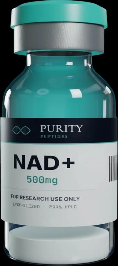
NAD+ consists of two nucleotides, one containing adenine and one containing nicotinamide, joined through their phosphate groups. The nicotinamide ring accepts a hydride ion to form NADH. A phosphorylated form, NADP+/NADPH, serves mainly in biosynthesis and antioxidant defence rather than energy production. NAD+ is not a peptide but is a common reagent in metabolic studies. In solution it is sensitive to pH and temperature, and NADH is light- and oxygen-sensitive.
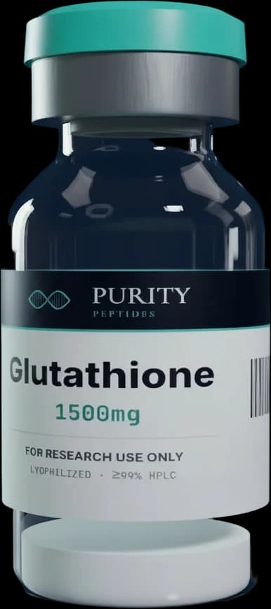
Glutathione is the tripeptide γ-L-glutamyl-L-cysteinyl-glycine. The γ-linkage between glutamate's side-chain carboxyl and cysteine makes it resistant to most peptidases. The cysteine thiol is the functional group: two reduced glutathione molecules (GSH) are oxidised to glutathione disulfide (GSSG). Intracellular concentrations are in the millimolar range, making glutathione the most abundant low-molecular-weight thiol in mammalian cells. In solution GSH oxidises readily, so handling and storage affect the GSH-to-GSSG ratio of research material.
Mechanisms studied
NAD+
- Energy metabolism. NAD+ accepts electrons in glycolysis and the tricarboxylic acid (TCA) pathway; NADH donates them to mitochondrial complex I to drive ATP synthesis.
- Sirtuins. The seven mammalian sirtuins (SIRT1–7) are NAD+-dependent deacylases that remove acetyl and other acyl groups from histones and metabolic enzymes, linking nutrient status to gene expression and mitochondrial function (Imai and Guarente, 2014).
- PARPs. Poly(ADP-ribose) polymerases consume NAD+ when repairing DNA damage. Heavy PARP activation can deplete cellular NAD+.
- CD38. This ectoenzyme is a major NAD+ consumer. Camacho-Pereira et al. (2016) reported that CD38 expression rises with age in mouse tissues and contributes to age-related NAD+ decline.
- Biosynthesis. Cells make NAD+ from tryptophan, from nicotinic acid, and through the salvage pathway from nicotinamide via NAMPT and the intermediate nicotinamide mononucleotide (NMN). Nicotinamide riboside (NR) feeds into the same pathway.
Glutathione
- Peroxide detoxification. Glutathione peroxidases use GSH to reduce hydrogen peroxide and lipid hydroperoxides, generating GSSG, which glutathione reductase recycles using NADPH. This is a direct biochemical link between the two molecules discussed here.
- Conjugation. Glutathione S-transferases attach glutathione to electrophilic compounds, a central route of xenobiotic detoxification.
- Protein regulation. Reversible S-glutathionylation of cysteine residues modulates protein function in response to oxidative conditions.
- Ferroptosis. The selenoenzyme GPX4 uses glutathione to reduce phospholipid hydroperoxides. Its inhibition, or depletion of glutathione, triggers ferroptosis, an iron-dependent form of regulated cell death (Dixon et al., 2012; Yang et al., 2014).
- Synthesis. Glutamate-cysteine ligase catalyses the rate-limiting step, with cysteine availability usually limiting (Lu, 2013).
Research and clinical-trial landscape
NAD+ and ageing. Many studies in rodents report that NAD+ levels decline in several tissues with age, and that raising NAD+ with precursors such as NMN or NR improves a range of metabolic and functional measures in aged or diet-challenged mice (Yoshino et al., 2011; Mills et al., 2016). Reviews by Verdin (2015), Rajman et al. (2018) and Covarrubias et al. (2021) summarise this body of work. Human research has so far consisted mainly of small randomised trials of NAD+ precursors that evaluated safety, blood NAD+ metabolome changes and selected physiological endpoints (for example Martens et al., 2018). These trials have generally confirmed that precursors raise NAD+-related metabolites in blood, but effects on clinical outcomes have been modest or inconsistent.
Glutathione and ageing. Lower glutathione levels and a more oxidised GSH-to-GSSG ratio have been reported in older individuals and in several chronic diseases. Research approaches include providing precursors such as cysteine sources and glycine (Sekhar et al., 2011) and studying glutathione itself (Richie et al., 2015). Because intact glutathione is extensively broken down and resynthesised, how well external glutathione raises intracellular levels in specific tissues remains debated.
Evidence quality and open questions
- Measurement challenges. NAD+ and glutathione are both unstable during sample handling. Differences in extraction and assay methods explain some inconsistencies between studies.
- Tissue specificity. Changes in blood levels may not reflect changes in muscle, liver or brain.
- Causality. Declining NAD+ or glutathione with age is well documented, but whether restoring them changes ageing biology in humans has not been shown in large, long-term trials.
- Double-edged biology. Raising NAD+ may also support proliferating or senescent cells, and lowering glutathione is studied as a way to sensitise tumour cells. Context matters.
Regulatory and anti-doping status
Neither NAD+ nor glutathione is an approved prescription medicine for ageing-related indications in the United States or EU. Both are sold as research reagents, and research-grade material is intended only for in vitro and animal research.
Neither substance is listed as prohibited by WADA. However, WADA's separate rules on prohibited methods (category M2) can apply regardless of the substance involved, so researchers working with athletes should consult the current list.
Summary table
| NAD+ | Glutathione | |
|---|---|---|
| Chemical class | Dinucleotide coenzyme | Tripeptide (γ-Glu-Cys-Gly) |
| Peptide? | No | Yes (γ-linked) |
| Redox pair | NAD+/NADH | GSH/GSSG |
| Key enzymes | Dehydrogenases, sirtuins, PARPs, CD38 | Glutathione peroxidases, reductase, GSTs, GPX4 |
| Ageing link studied | Age-related tissue decline; CD38 | Lower GSH and more oxidised ratio with age |
| Human evidence | Small precursor trials | Small precursor and glutathione studies |
| Status | Research reagent; not an approved drug | Research reagent; not an approved drug |
| WADA | Not listed; M2 method rules apply | Not listed; M2 method rules apply |
References
- Verdin E. NAD+ in aging, metabolism, and neurodegeneration. Science. 2015;350(6265):1208–1213.
- Rajman L, Chwalek K, Sinclair DA. Therapeutic potential of NAD-boosting molecules: the in vivo evidence. Cell Metab. 2018;27(3):529–547.
- Covarrubias AJ, Perrone R, Grozio A, Verdin E. NAD+ metabolism and its roles in cellular processes during ageing. Nat Rev Mol Cell Biol. 2021;22(2):119–141.
- Imai S, Guarente L. NAD+ and sirtuins in aging and disease. Trends Cell Biol. 2014;24(8):464–471.
- Camacho-Pereira J, Tarragó MG, Chini CCS, et al. CD38 dictates age-related NAD decline and mitochondrial dysfunction through an SIRT3-dependent mechanism. Cell Metab. 2016;23(6):1127–1139.
- Yoshino J, Mills KF, Yoon MJ, Imai S. Nicotinamide mononucleotide, a key NAD+ intermediate, treats the pathophysiology of diet- and age-induced diabetes in mice. Cell Metab. 2011;14(4):528–536.
- Mills KF, Yoshida S, Stein LR, et al. Long-term administration of nicotinamide mononucleotide mitigates age-associated physiological decline in mice. Cell Metab. 2016;24(6):795–806.
- Martens CR, Denman BA, Mazzo MR, et al. Chronic nicotinamide riboside supplementation is well-tolerated and elevates NAD+ in healthy middle-aged and older adults. Nat Commun. 2018;9:1286.
- Meister A, Anderson ME. Glutathione. Annu Rev Biochem. 1983;52:711–760.
- Lu SC. Glutathione synthesis. Biochim Biophys Acta. 2013;1830(5):3143–3153.
- Forman HJ, Zhang H, Rinna A. Glutathione: overview of its protective roles, measurement, and biosynthesis. Mol Aspects Med. 2009;30(1–2):1–12.
- Sekhar RV, Patel SG, Guthikonda AP, et al. Deficient synthesis of glutathione underlies oxidative stress in aging and can be corrected by dietary cysteine and glycine supplementation. Am J Clin Nutr. 2011;94(3):847–853.
- Richie JP Jr, Nichenametla S, Neidig W, et al. Randomized controlled trial of oral glutathione supplementation on body stores of glutathione. Eur J Nutr. 2015;54(2):251–263.
- Dixon SJ, Lemberg KM, Hayano M, et al. Ferroptosis: an iron-dependent form of nonapoptotic cell death. Cell. 2012;149(5):1060–1072.
- Yang WS, SriRamaswamy R, Viswanathan VS, et al. Regulation of ferroptotic cancer cell death by GPX4. Cell. 2014;156(1–2):317–331.
FOR RESEARCH USE ONLY.Not for human or veterinary use. Not intended to diagnose, treat, cure, or prevent any disease.